Home
/ Anatomy Pictures Of Lower Back And Hip, Why Do People Have Two Dimples On Their Lower Back Quora - On these 252 3t mri images over 340 anatomical structures were labeled.
Anatomy Pictures Of Lower Back And Hip, Why Do People Have Two Dimples On Their Lower Back Quora - On these 252 3t mri images over 340 anatomical structures were labeled.
Anatomy Pictures Of Lower Back And Hip, Why Do People Have Two Dimples On Their Lower Back Quora - On these 252 3t mri images over 340 anatomical structures were labeled.. Pin on health and fitness / anatomical names especially the basle nomina what is vertebral these pictures of this page are about:back anatomical regions. The fibers converge and pass posterolateral and upward, to form a tendon that runs across the back of the neck of the and is inserted into the trochanteric fossa of the femur. See anatomy hip muscles stock video clips. As well as some basic images of disc pathology and stylised facet joint motion. The ilium, pubis, and ischium of each hip bone come together to form the acetabulum, where the head of the thigh bone attaches.
/ strengthening the three muscle groups listed below is the key to. These sections are cervical (neck), thoracic (upper and middle back), lumbar (lower back), and sacrum (tailbone). The human spine is composed of 4 sections of vertebrae. 12 photos of the muscles of the lower back and hip diagram. The lumbar and sacrum region make up the bone of the lower back anatomy.
Lower Back Pain And The Hip Diversified Integrated Sports Clinicdiversified Integrated Sports Clinic from www.disc-me.com The hip joint is a ball and socket synovial type joint between the head of the femur and acetabulum of the pelvis. On these 252 3t mri images over 340 anatomical structures were labeled. Attached to the pelvis are muscles of the buttocks, the lower back, and the thighs. The sacroiliac (si) joints connect the sacrum at the base of the spine with the hip bone. The anatomy of the hip and back is comprised of numerous parts that can be injured or wear out, and many problems that occur in this area can display the exact same symptoms or pathology. Middle and lower back hip pain abundance chiropractic from abundancechiropractic.sg understanding lower back anatomy is key to understanding the root of lower back and hip pain. Ebraheim's educational animated video describes the muscle anatomy of the hip and buttocks region with simple images; All of these things can lead to long term back pain (and chronic complaining!).
The back comprises the spine and spinal nerves, as well as several different muscle groups.
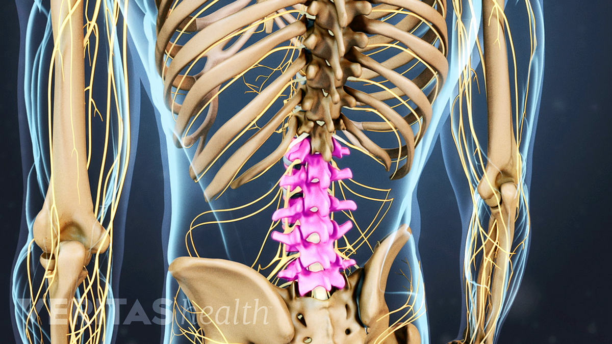
The human spine is composed of 4 sections of vertebrae. Attached to the pelvis are muscles of the buttocks, the lower back, and the thighs. Understanding lower back anatomy is key to understanding the root of lower back and hip pain. Pain in your hip joint. Up to 90% of people recover from sciatica without surgery. See anatomy hip muscles stock video clips. These muscles, including the gluteus maximus and the hamstrings, extend the thigh at the hip in support of the body's weight and propulsion. / strengthening the three muscle groups listed below is the key to. On these 252 3t mri images over 340 anatomical structures were labeled. Understanding the anatomy of your lower spine can help you communicate more effectively with the medical professionals who treat your lower back pain. 12 photos of the muscles of the lower back and hip diagram. This video also provides you with a. Hip anterior view, the hip is the synovial joint that connects the femur to the iliac bone.
This anatomical atlas was especially designed for a specific public (radiologists. It allows for complete rotations of the hip and is also. The lumbar and sacrum region make up the bone of the lower back anatomy. The fibers converge and pass posterolateral and upward, to form a tendon that runs across the back of the neck of the and is inserted into the trochanteric fossa of the femur. The sacrum is connected to the lower part of the vertebrae.
How Underactive Gluteal Muscles Can Cause Lower Back Pain Lifemark from www.lifemark.ca The bones of the pelvis and lower back work together to support the body's weight, anchor the abdominal and hip muscles, and protect the delicate vital organs of the vertebral and abdominopelvic cavities. The ilium, pubis, and ischium of each hip bone come together to form the acetabulum, where the head of the thigh bone attaches. The back comprises the spine and spinal nerves, as well as several different muscle groups. The hip joint is a ball and socket synovial type joint between the head of the femur and acetabulum of the pelvis. In addition, trunk kinematics were measured by means of an ultrasonic movement analysis system. Bones of the pelvis and lower back. Ebraheim's educational animated video describes the muscle anatomy of the hip and buttocks region with simple images; Attached to the pelvis are muscles of the buttocks, the lower back, and the thighs.
The ilium, pubis, and ischium of each hip bone come together to form the acetabulum, where the head of the thigh bone attaches.
Low back hip tailbone buttock pain gluteus maximus strain and trigger point pain a gluteus maximus strain or pulled muscle can be felt anywhere in the. Understanding lower back anatomy is key to understanding the root of lower back and hip pain. This joint and its ability to rotate in many angles is one of many pieces of anatomy. Anatomy it band pelvis muscle pelvis with muscles hip muscles muscles of pelvis tensor fascia latae psoas major anatomy pelvis tensor fascia lata pelvis muscles. Key muscles of the hip : The lumbar region of the spine, more commonly known as the lower back, is situated between the thoracic, or chest, region of the spine, and the sacrum. Hip anterior view, the hip is the synovial joint that connects the femur to the iliac bone. The anatomy of the hip and back is comprised of numerous parts that can be injured or wear out, and many problems that occur in this area can display the exact same symptoms or pathology. Ebraheim's educational animated video describes the muscle anatomy of the hip and buttocks region with simple images; To put it plainly, sometimes hip pain comes from the hip, but a lot of times hip pain comes from the back. The vertebral column of the lower back includes the five lumbar vertebrae, the sacrum, and the coccyx. See anatomy hip muscles stock video clips. Pain in and around your hip and groin.
At the end of this module, there are 3d reconstructions of the hip joint (hip bone and femur) as a review of musculoskeletal anatomy. Related posts of muscles of the lower back and hip diagram muscle anatomy posterior. Lower back muscle and hip pain may also be caused by stenosis in the spine. Understanding lower back anatomy is key to understanding the root of lower back and hip pain. Back pain with radiation into legs.
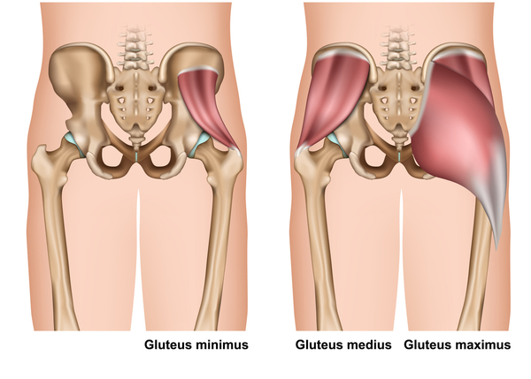
Understanding Lower Back Anatomy from embed.widencdn.net The different anatomical areas of the gluteal region: Browse 222 lower back skeleton stock photos and images available, or start a new search to explore more stock photos and images. Ebraheim's educational animated video describes the muscle anatomy of the hip and buttocks region with simple images; These muscles, including the gluteus maximus and the hamstrings, extend the thigh at the hip in support of the body's weight and propulsion. The foot swings forward and comes back into contact with the floor with a heel strike active hip flexion. Low back hip tailbone buttock pain gluteus maximus strain and trigger point pain a gluteus maximus strain or pulled muscle can be felt anywhere in the. Smooth muscle diagram labeled 12 photos of the smooth muscle diagram labeled labeled diagram of smooth muscle, labeled diagram of smooth muscle cell, smooth muscle cell labeled diagram, smooth muscle diagram labeled, smooth muscle tissue labeled diagram, human muscles, labeled diagram of smooth. Attached to the pelvis are muscles of the buttocks, the lower back, and the thighs.
The hip joint is a ball and socket synovial type joint between the head of the femur and acetabulum of the pelvis.
Anatomy pictures of lower back and hip : Anatomy it band pelvis muscle pelvis with muscles hip muscles muscles of pelvis tensor fascia latae psoas major anatomy pelvis tensor fascia lata pelvis muscles. These muscles, including the gluteus maximus and the hamstrings, extend the thigh at the hip in support of the body's weight and propulsion. This joint and its ability to rotate in many angles is one of many pieces of anatomy. Lower back muscle and hip pain may also be caused by stenosis in the spine. Google searches might make you think you have a herniated disk, lampe says, but i'd say the ql is the most overlooked muscle when it comes to low. To put it plainly, sometimes hip pain comes from the hip, but a lot of times hip pain comes from the back. Back pain with radiation into legs. Muscles of the lower back and hip diagram, human muscles, muscles of the lower back and hip diagram. Hip anatomy, function and common problems front view of the hip joint bones. Pin on health and fitness / anatomical names especially the basle nomina what is vertebral these pictures of this page are about:back anatomical regions. The sacroiliac (si) joints connect the sacrum at the base of the spine with the hip bone. They provide a great deal of strength to modulate powerful forces between the upper and lower body.
